[:de]
Assistierte Reproduktion, also medizinische Nachhilfe bei der Fortpflanzung, kommt im Artenschutz von extrem seltenen Tieren wie Spix-Ara oder nördlichen Breitmaulnashörnern in den letzten Jahren immer häufiger zum Zuge. Bei Reptilien gibt es bisher dagegen erst wenige Studien zur assistierten Reproduktion, speziell bei Chamäleons nur vereinzelte. Wissenschaftler aus den USA haben nun eine Studie dazu an männlichen Jemen- und Pantherchamäleons (Chamaeleo calyptratus und Furcifer pardalis) durchgeführt.
An der Louisiana State University wurden je 16 Männchen beider Arten unter standardisierten Bedingungen über ein Jahr lang gehalten. Die Pantherchamäleons wurden bei einem US-amerikanischen Züchter erworben, die Jemenchamäleons von einem Händler, der sie wiederum der Population wild lebender Jemenchamäleons in Florida entnommen hatte. Alle Männchen wurden einzeln in ReptiBreeze gehalten, ausgestattet mit automatischer Beregnung und künstlichen Pflanzen. Die Temperaturen lagen tagsüber bei rund 28-29°C mit Spots zum Aufsuchen höherer Werte. 12 h UV-B-Bestrahlung am Tag wurde angeboten. Gefüttert wurde mit Heimchen und Zophobas.
Vor Beginn der Studie wurden alle 32 Chamäleons klinisch untersucht und mehrere Parasitenbehandlungen durchgeführt. Erst nach einem Monat der Akklimatisierung begann dann die eigentliche Studie. Während des Studienjahres wurden alle Chamäleons zwei Mal pro Monat in Narkose gelegt. Jedes Mal wurde Blut aus der ventralen Schwanzvene oder der Jugularvene entnommen, um die Testosteron-Konzentration zu bestimmen. Mittels Ultraschalles wurde die Größe der Hoden vermessen. Zudem wurde jedes Mal versucht, mittels Elektroejakulation Sperma zu gewinnen. Bei der Elektroejakulation wurde eine kleine Metallsonde in die gesäuberte Kloake eingeführt. Jedes Chamäleon wurde dann bis zu drei Mal hintereinander mit bis zu 15 Stromstößen von 0,1/0,2/0,3 mAs behandelt. Die Absamversuche wurden abgebrochen, sobald das Tier ejakulierte. Das gewonnene Sperma wurde konserviert und auf Ejakulatvolumen, Vorhandensein von Spermien, Spermienbeweglichkeit, -konzentration und -morphologie untersucht.
Die Ergebnisse lassen vermuten, dass Jemenchamäleons bei gleichbleibenden Haltungsbedingungen eine sogenannte prenuptiale Fortpflanzungsstrategie verfolgen. Die Testosteronkonzentration im Blut stieg bereits an, bevor das Spermavolumen der Männchen ihr Maximum erreicht hatte. Die besten Absamerfolge brachten die Monate Mai, April und Juni, das meiste Sperma brachten Elektroejakulationen im dritten Anlauf. Auch die Hodengrößen unterschieden sich übers Jahr, mit den größten Messungen von August bis Dezember.
Pantherchamäleons dagegen scheinen einer postnuptialen Fortpflanzungsstrategie zu folgen. Bei ihnen konnte das meiste Sperma erst weit nach dem höchsten Punkt der Testosteronkonzentration gewonnen werden. Die Absamungen klappten am besten im März, April, Mai und Juni. Wesentlich häufiger als bei Jemenchamäleons funktionierte die Elektroejakulation bei Pantherchamäleons schon im ersten Versuch. Die Hodengrößen variierten auch hier übers Jahr, allerdings waren sie mehrheitlich in den schon genannten Monaten am größten. Gemeinsam mit den genannten Faktoren veränderte sich ebenso das Volumen des Ejakulats, die Spermienkonzentration, -beweglichkeit und -morphologie im Jahresverlauf.
Die Autoren empfehlen, Elektroejakulation bei Chamäleons generell nur in Narkose durchzuführen. Die Erfolgsrate beim Absamen lag in den beiden höchsten Fällen bei 82 und 88%, was den Erfolgen bei anderen Reptilien während deren Fortpflanzungssaison entspricht. Die Mortalitätsrate unter den 32 Tieren lag über das ganze Jahr lediglich bei 0,12%. Ein Pantherchamäleon starb nach 10 Monaten während der 20. Narkose, nach dem Tod wurde ein Nierenschaden festgestellt. Aus der geringen Mortalitätsrate schließen die Autoren, dass die Elektroejakuation eher keine Rolle in der Entwicklung von Nierenerkrankungen spiele, wie es in anderen Studien vermutet wurde. Eine Untersuchung des Bluts auf Nierenwerte wurde allerdings bei keinem der überlebenden Chamäleons nach der Studie durchgeführt. Unklar bleibt auch, welche Rolle die fehlende Imitation von Regen- und Trockenzeit im Jahresverlauf für beide Arten und deren Fortpflanzungszyklus spielt.
Characterizing the annual reproductive cycles of captive male veiled chameleons (Chamaeleo calyptratus) and panther chameleons (Furcifer pardalis)
Sean M. Perry, Sarah R. Camlic, Ian Konsker, Michael Lierz, Mark A. Mitchell
Journal of Herpetological Medicine and Surgery 33 (1), 2023, pp. 45-60
DOI: 10.5818/JHMS-D-22-00037
[:en]
Assisted reproduction has become increasingly common in the conservation of extremely rare animals such as the Spix’s macaw or northern white rhinoceros in recent years. In reptiles, on the other hand, there have only been a few studies on assisted reproduction, and only a few on chameleons in particular. Scientists from the USA have now conducted a study on male Veiled and Panther Chameleons (Chamaeleo calyptratus and Furcifer pardalis).
At Louisiana State University, 16 males of each species were kept under standardised conditions for over a year. The panther chameleons were purchased from a US breeder, the Yemen chameleons from a dealer who had taken them from the introduced wild chameleon population in Florida. All males were kept individually in ZooMed screen cages, equipped with automatic sprinklers and artificial plants. Temperatures were around 28-29°C during the day with spots to seek higher values. 12 h UV-B irradiation per day was offered. They were fed with crickets and zophobas.
Before the start of the study, all 32 chameleons were clinically examined and parasites were treated. Only after a month of acclimatisation did the actual study begin. During the study year, all chameleons were put under anaesthesia twice a month. Each time, blood was taken from the ventral tail vein or the jugular vein to determine the testosterone concentration. Ultrasound was used to measure the size of the testicles. In addition, each time an attempt was made to obtain sperm by electroejaculation. Electroejaculation involved inserting a small metal probe into the cleaned cloaca. Each chameleon was then treated up to three times in succession with up to 15 electric shocks of 0.1/0.2/0.3 mAs. The semen collection experiments were stopped as soon as the animal ejaculated. The sperm collected was preserved and examined for ejaculate volume, presence of sperm, sperm motility, concentration, and morphology.
The results suggest that Veiled Chameleons follow a so-called prenuptial reproductive strategy under constant husbandry conditions. The testosterone concentration in the blood already increased before the sperm volume of the males had reached its maximum. The months of May, April, and June brought the best sperm volumes, the most sperm was produced by electroejaculations in the third attempt. Testicle sizes also varied throughout the year, with the largest measurements from August to December.
Panther chameleons, on the other hand, seem to follow a postnuptial reproductive strategy. In them, most sperm could only be obtained well after the highest point of testosterone concentration. The electroejaculations worked best in March, April, May and June. Much more often than in Yemen chameleons, electroejaculation in panther chameleons worked already in the first attempt. The size of the testicles also varied throughout the year, but most were largest in the months mentioned above. Together with the factors mentioned above, the volume of ejaculate, sperm concentration, sperm motility and sperm morphology also changed during the year.
The authors recommend that electroejaculation in chameleons should generally only be performed under anaesthesia. The success rate for spermatozoa in the two highest cases was 82 and 88%, which is similar to the success in other reptiles during their reproductive season. The mortality rate among the 32 animals was only 0.12% over the whole year. One panther chameleon died after 10 months during the 20th anaesthesia, after death kidney damage was detected. From the low mortality rate, the authors conclude that electroejaculation rather does not play a role in the development of kidney disease, as was suspected in other studies. However, an examination of the blood for kidney values was not carried out on any of the surviving chameleons after the study. It also remains unclear what role the lack of imitation of rainy and dry seasons during the year plays for both species and their reproductive cycle.
Characterizing the annual reproductive cycles of captive male veiled chameleons (Chamaeleo calyptratus) and panther chameleons (Furcifer pardalis)
Sean M. Perry, Sarah R. Camlic, Ian Konsker, Michael Lierz, Mark A. Mitchell
Journal of Herpetological Medicine and Surgery 33 (1), 2023, pp. 45-60
DOI: 10.5818/JHMS-D-22-00037
[:]
![[:de]Neue Fallberichte zur Hemipenesamputation[:en]New case reports on hemipenes amputation[:]](https://www.agchamaeleons.de/wp-content/uploads/2024/04/Furcifer-pardalis-Lokalform-Ambilobe-Foto-Alex-Laube-4.jpg "[:de]Neue Fallberichte zur Hemipenesamputation[:en]New case reports on hemipenes amputation[:]")
![[:de]Hautverfärbungen nach Mückenstichen[:en]Mosquito bites may induce skin colour change[:]](https://www.agchamaeleons.de/wp-content/uploads/2024/02/Mosquito-bites-in-chameleons.jpg "[:de]Hautverfärbungen nach Mückenstichen[:en]Mosquito bites may induce skin colour change[:]")
![[:de]Histologie der Chamäleonleber[:en]Histology of the chameleon liver[:]](https://www.agchamaeleons.de/wp-content/uploads/2023/11/Histo-Jemenchamaeleon-Leber.jpg "[:de]Histologie der Chamäleonleber[:en]Histology of the chameleon liver[:]")
![[:de]Jemenchamäleon sucht neue Halter[:en]Veiled Chameleon seeks new keeper[:]](https://www.agchamaeleons.de/wp-content/uploads/2023/07/Chamaeleo-calyptratus-public-domain-1-784x606.jpg "[:de]Jemenchamäleon sucht neue Halter[:en]Veiled Chameleon seeks new keeper[:]")
![[:de]Zoonotisches Potenzial von Jemenchamäleons auf Gran Canaria (Spanien)[:en]Zoonotic potential of Yemen chameleons in Gran Canaria (Spain)[:]](https://www.agchamaeleons.de/wp-content/uploads/2023/07/Chamaeleo-calyptratus-CC-3.0-Eureus-verkleinert.jpg "[:de]Zoonotisches Potenzial von Jemenchamäleons auf Gran Canaria (Spanien)[:en]Zoonotic potential of Yemen chameleons in Gran Canaria (Spain)[:]")
![[:de]Zwillingsschlupf bei Jemenchamäleons in Lettland[:en]Twins in Veiled Chameleons in Latvia[:]](https://www.agchamaeleons.de/wp-content/uploads/2023/07/Zwillinge-Jemenchams.jpg "[:de]Zwillingsschlupf bei Jemenchamäleons in Lettland[:en]Twins in Veiled Chameleons in Latvia[:]")
![[:de]Wie und wann Jemenchamäleons vibrieren[:en]How and when Veiled Chameleons show biotremors[:]](https://www.agchamaeleons.de/wp-content/uploads/2023/07/Chamaeleo-calyptratus-public-domain-1.jpg "[:de]Wie und wann Jemenchamäleons vibrieren[:en]How and when Veiled Chameleons show biotremors[:]")
![[:de]Langzeitstudie zur Spermagewinnung bei Chamäleons[:en]Long-term study on sperm collection in chameleons[:]](https://www.agchamaeleons.de/wp-content/uploads/2023/03/Furcifer-pardalis-Foto-Alex-Laube.jpg "[:de]Langzeitstudie zur Spermagewinnung bei Chamäleons[:en]Long-term study on sperm collection in chameleons[:]")
![[:de]Vergleiche an Gliedmaßen von Hadramautagame und Jemenchamäleon[:en]Comparisons on limbs of Hadramaut agama and Veiled Chameleon[:]](https://www.agchamaeleons.de/wp-content/uploads/2023/03/Chamaeleo-calyptratus-CC-3.0-Kupos-verkleinert.jpg "[:de]Vergleiche an Gliedmaßen von Hadramautagame und Jemenchamäleon[:en]Comparisons on limbs of Hadramaut agama and Veiled Chameleon[:]")
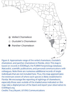
![[:de]Eingeschleppte Chamäleons in Florida[:en]Introduced chameleons in Florida[:]](https://www.agchamaeleons.de/wp-content/uploads/2023/01/Furcifer-pardalis-Ambilobe-male-2019-3.jpg "[:de]Eingeschleppte Chamäleons in Florida[:en]Introduced chameleons in Florida[:]")