Lecture for vets on prophylaxis for chameleons

Lecture for vets on prophylaxis for chameleons

Tiermedizin Webinars

On 18 November 2023, the spokesperson of the AG Chameleons will give a lecture for veterinary colleagues on which prophylactic measures are possible and useful in veterinary practice. To a large extent, prophylaxis includes keeping the chameleons in the terrarium itself, so a large part of the lecture will deal with what needs to be considered when keeping chameleons and which common mistakes still lead to husbandry-related diseases or injuries in chameleons. She will also discuss the collection of the so-called minimum database during the annual check-up in the veterinary practice and show examples of diseases recognised early and late. A Q&A session directly after the respective presentations rounds off the short excursion into prophylaxis in chameleons.

Registration for the conference is also possible for non-DGHT members who are veterinarians – see the link below to the mVet conference platform.

Dr. Alexandra Laube Prophylaxis for chameleons – is it possible and if so, how?
59. Conference of DGHT working group amphibian and reptile diseases (AG ARK)
Online

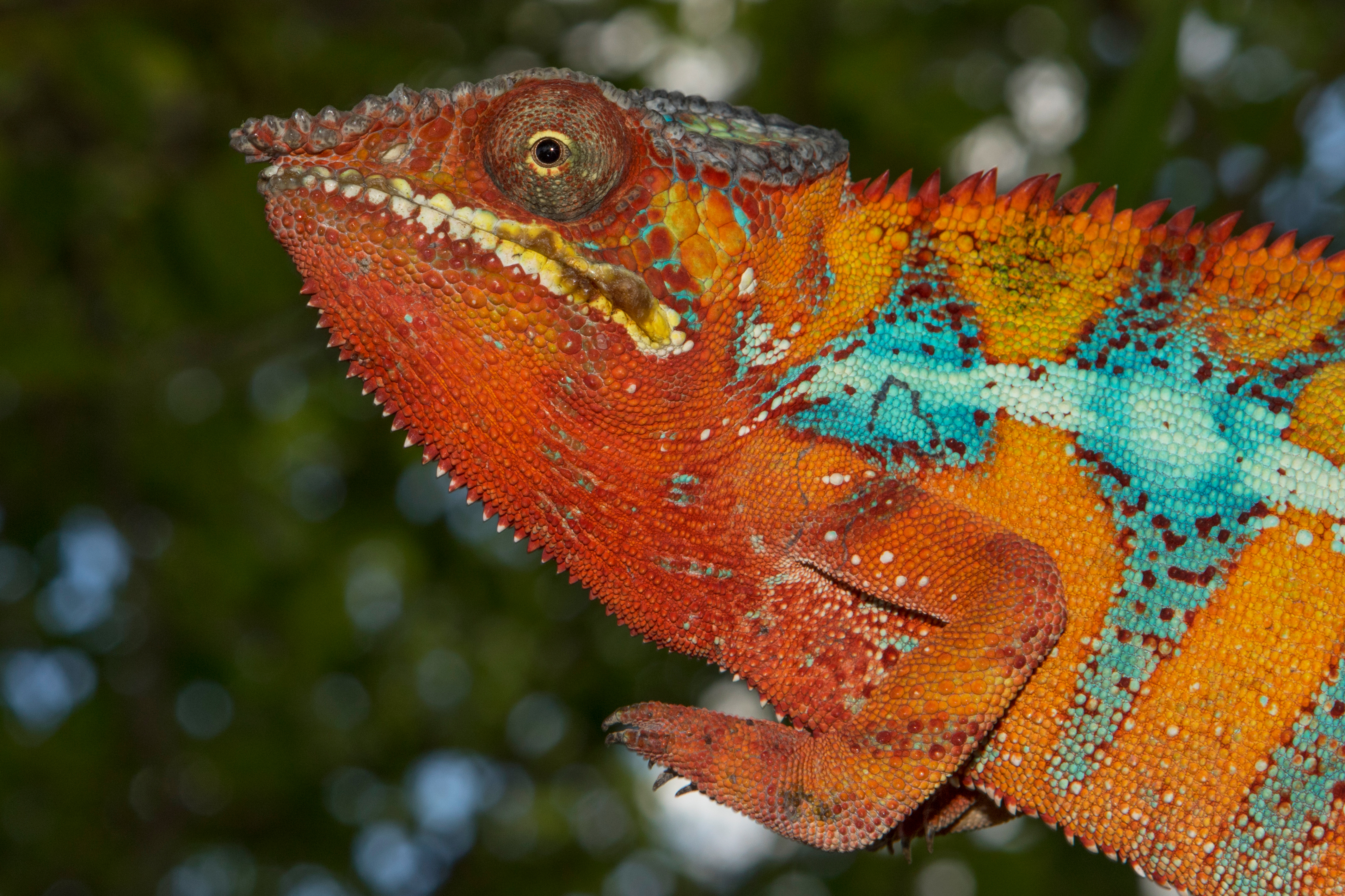
Photo: Calumma amber in the Montagne d’Ambre, Madagascar, photographed by A. Laube

Histology of the chameleon liver

Histology of the chameleon liver

Tiermedizin Science

Histological examinations of organ tissue are part of every pathological examination in veterinary medicine. They are also frequently carried out in reptiles, but there are few studies on the histology of healthy organ tissue. An Arabic publication now deals with histological sections of chameleon livers.

Seven adult Yemen chameleons were captured in Abha City in the Aseer region and then killed with ether inhalation. The livers were placed in formalin and then poured into paraffin to make sections.

Morphologically, the liver was found to be a two-lobed, dark brown organ approximately 3.7 x 2 cm in size, which lies in the coelomic cavity in front of the stomach and surrounds the gall bladder. As in other animals, a capsule of connective tissue surrounds the liver.

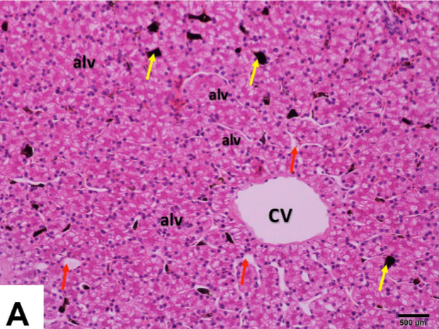
Histologically, the liver of Yemen chameleons resembles that of other vertebrates in many respects. The liver capsule consists of closely spaced collagenous fibres and smooth muscle fibres. Normally, trabecular connective tissue divides the liver itself into many small lobules, but such a structure does not appear to be present in Yemen chameleons. In contrast to mammals, the liver cells (hepatocytes) are not arranged radially around a vein, but rather irregularly in follicles or alveoli. The hepatocytes are surrounded by capillary blood vessels. So-called melanoma macrophages, which are not found in birds and mammals, can be seen in the blood vessels. The hepatocytes in the Yemen chameleon are polyhedral or pyramid-shaped and usually contain several large, round cell nuclei in the periphery. The nuclei contain conspicuously dark nucleoli. Occasionally nuclei are central. Under haematoxylin-eosin (HE) staining, the hepatocytes appear very eosinophilic. In the connective tissue, branches of the portal vein, hepatic artery, small bile ducts and lymphatic vessels could be visualised. Haematopoietic tissue was found in the area directly under the liver capsule.

In addition to the histological examination, several pieces of liver were also examined using transmission electron microscopy. Images of both examination methods can be found in the publication.

Histomorphological, histochemical and ultrastructural studies on the healthy liver of Yemen Veiled Chameleon (Chamaeleo calyptratus) in Southern Saudi Arabia
Amin A. Al-Doaiss, Mohammed A. Alshehri, Ali A. Shati, Mohammad Y. Alfaifi, Mohammed A. Al-Kahtani, Ahmed Ezzat Ahmed, Refaat A. Eid, Laila A. Al-Shuraym, Fahd A. Al-Mekhlafi, Mohammed Al Zahrani, Mohammed Mubarak
International Journal of Morphology 41(5), 2023: pp. 1513-1526.
DOI: none

Image: Histological section of the liver of a Yemen chameleon from the above-mentioned publication

Effect of human chorionic gonadotropin in chameleons

Effect of human chorionic gonadotropin in chameleons

Tiermedizin Science

After a study on sperm collection in chameleons was already published this year, further results from the largely same team of authors now follow. The aim is to further research the basics of assisted reproduction, i.e. medical assistance in reproduction, in chameleons.

At Louisiana State University, 24 Veiled Chameleons were kept under standardised conditions for over a year. All animals came from a dealer who had taken them from the population of wild Veiled Chameleons in Florida. All were kept individually in ReptiBreeze, equipped with automatic sprinklers and artificial plants. Temperatures were around 28-29°C during the day with spots to seek higher values. 12 h UV-B irradiation per day was offered. Crickets and zophobas were fed. Before the start of the study, all 24 chameleons were clinically examined and several parasite treatments were carried out. Only after a month of acclimatisation did the actual study begin.

The first experiment tested what dose of human choriogonadotropin (hCG) is needed to increase the hormone levels of testosterone in the blood by 50%. Eleven Veiled Chameleons were randomly assigned to one of three groups. The three groups received injections of 100, 200 or 300 IU hCG under the skin at two-week intervals. Blood samples were taken before the first hormone injection and at 30 minutes, one hour, two hours, four hours, eight hours, 12 hours and 24 hours afterwards.

The second experiment tested the effect of hCG treatment on sperm production. 13 Veiled Chameleons were randomly assigned to a treatment or a control group. Once a week for one month, the animals in the first group were treated with 100 IU of hCG, while the second group was only injected with the same volume of isotonic saline. After a four-week break, the groups were switched and the experiment repeated. Blood samples to measure testosterone levels were taken before treatment and on day 15 and 30 afterwards. Semen was collected by electroejaculation under anaesthesia on the day before treatment and 30 days after.

The results showed that the testosterone level in male Veiled Chameleons increased significantly directly after the administration of hCG and remained elevated for about 24 hours. However, it did not matter which dose of hCG had been given beforehand. It could also be shown that the testosterone level increased significantly after the administration of hCG compared to the control group, which only received saline solution. The number of successful electroejaculations could be increased under hCG.

Effects of exogenous human chorionic gonadotropin administration on plasma testosterone and semen production in the Veiled Chameleon (Chamaeleo calyptratus)
Sean M. Perry, Sarah R. Camlic, Michael Lierz, Mark A. Mitchell
Journal of Herpetological Medicine and Surgery 33 (3), 2023, pp. 180-191
DOI: 10.5818/JHMS-D-22-00038

The microbiome of dwarf chameleons

The microbiome of dwarf chameleons

Tiermedizin Science

The term microbiome has been very popular for some years now. In humans and animals, it refers to the totality of all microorganisms that colonise a living being. Most of them colonise the gastrointestinal tract. In the case of chameleons, there is only very limited literature on this topic. A master’s thesis from South Africa now deals with the bacterial composition of the microbiome in South African dwarf chameleons of the genus Bradypodion.

60 cheek swabs were collected from wild chameleons in KwaZulu-Natal. Of these, 20 were cheek swabs from Bradypodion melanocephalum, 20 from Bradypodion thamnobates and 20 from Bradypodion setaroi. After sampling, the same 60 animals were transported in cloth bags to the research base, where the animals were kept in 3.3 l boxes for 24 hours to obtain faecal samples. Since not all of the original 60 chameleons defecated, faeces were collected from additional chameleons.

The samples were all genetically tested. 40.43% of the samples contained Firmicutes, a similarly large proportion of the samples contained Proteobacteria with 36.86%. Bacteroidota followed with some distance, which could be detected in just under 16% of the samples. Verrucomicrobiota, Fusobacteriota, Actinobateriota, Spirochetes, Desulfobacteroa, Cyanobacteria, Thermoplamatota, Deferribacterota, Synergistota, Campylobacterota, Deinococcota, Halobacterota, Euryarchaeota, Elusimicrobiota and Myxococcota were found in significantly smaller numbers (up to 2%).

The microbiome of dwarf chameleons of the species Bradypodion melanocephalum, Bradypodion thamnobates and Bradypodion setaroi is similar to that of other reptiles. It consists mainly of proteobacteria and firmicutes, which may contribute to digestion. One particular bacterial species also suggests that the diet of the studied dwarf chameleons may include beetles of the genus Dendrophagus. The microbiome of all three dwarf chameleon species was very similar in the cheek swabs – this is called phylosymbiosis – while there were differences in composition between the species in the faeces. In all three dwarf chameleon species, significantly more different bacteria were found in the faeces than in the cheek swabs. A comparison between males and females did not reveal any significant differences in the microbiome of all three chameleon species. The author assumes that the bacterial species depend on the different habitats of the respective species. It is still unclear to what extent the microbiome is related to bacteria that a chameleon may ingest with feeding insects or from the soil of its environment. A detailed list of the bacterial species found can be found in the appendix of the publication.

The Hitchhiker’s Guide to dwarf chameleons (Bradypodion): The composition and function of the microbiome
Matthew G. Adair
Master of Science dissertation at the university of Johannesburg, 2023
DOI: not available

Comparative anatomy of the forearms of different chameleons

Comparative anatomy of the forearms of different chameleons

Tiermedizin Science

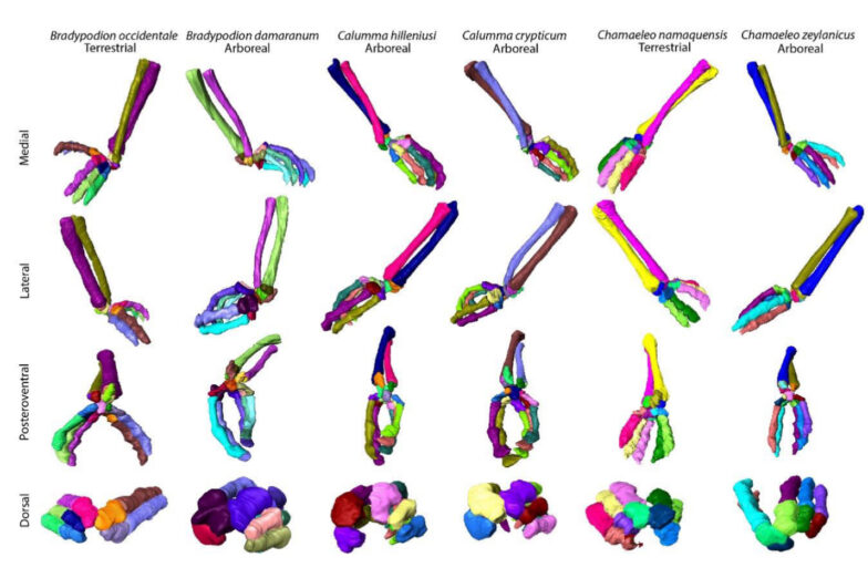
The anatomy of chameleons seems to be strongly adapted to their way of life. Tree-dwellers differ in many aspects from ground-dwellers. Several studies at the University of South Dakota this year have already looked at various anatomical aspects of chameleons. A new study is dedicated to the hands and arms.

For the investigation, the arms and hands of a total of 12 chameleons were isolated from existing microcomputer tomography scans and displayed in 3D. These were measured to about 30 different lengths and widths using software. Scans of the species Bradypodion damaranum, Bradypodion occidentale, Calumma hilleniusi, Calumma crypticum, Chamaeleo namaquensis, Chamaeleo zeylanicus, Furcifer balteatus, Furcifer campani, Rhampholeon spinosus, Rhampholeon temporalis, Trioceros goetzei goetzei and Trioceros werneri were evaluated. When selecting the species, care was taken to select one strictly tree-inhabiting chameleon and one more ground-inhabiting chameleon per genus.

The evaluation revealed that tree-dwelling chameleons show few differences in forelimb anatomy compared to ground-dwelling chameleons. Tree-dwelling species showed a majority of separated metacarpal bones 1-3, while ground-dwelling species showed fused metacarpal bones. Interestingly, this study differs from earlier studies by other authors, which yielded different results. The relatively small number of animals studied may be related to this. Larger studies could be helpful here.

Ecological and evolutionary drivers of chameleon forelimb variation
Ellie M. Schley
Honors Thesis 302 der Universität von South Dakota, 2023
DOI: gibt es nicht

Aggressive fungal pathogen discovered in panther chameleons

Aggressive fungal pathogen discovered in panther chameleons

Tiermedizin Science

Fungi of the genera Nannizziopsis and Paranannizziopsis have long been known to cause severe skin diseases in various reptiles. These include species feared in herpetocolture such as CANV (Chrysosporium Anamorph of Nannizziopsis vriesii) and Nannizziopsis dermatitidis, which are apparently obligate pathogens. Now, a similar skin fungus has been detected in panther chameleons in Florida, USA.

Nine adult panther chameleons (Furcifer pardalis) were taken from a wild population in Florida. They were first housed in groups of two or three chameleons in screen cages with natural and artificial plants at a private keeper. A ReptiSun 5.0 lamp and a conventional light bulb were used. The animals were fed with crickets and zophobas every second day and supplemented with vitamins and calcium. All nine panther chameleons, plus a tenth that was captured later, were finally given to the United States Department of Agriculture (USDA) for a series of experiments. They were kept individually in steel aviaries outdoors.

Subsequently, the eight remaining panther chameleons were also examined. In fact, all but one of the chameleons were found to have either missing claws or swelling of the hands and feet, small skin wounds, circumferential proliferation on the body and/or yellow and black skin lesions. Fungal PCR was no longer carried out, but infection with the same pathogen was suspected. All nine panther chameleons still alive were treated with 25 mg/kg terbinafine and 5 mg/kg voriconazole, both given orally once daily.

After six weeks, the panther chameleons were examined again. The skin lesions were still present, in two animals the hand and foot swellings had decreased. After eleven to twelve weeks of treatment, all symptoms had disappeared in seven chameleons. The skin lesions had developed into scars. Only two chameleons still showed swelling in the foot area, but less than at the beginning of treatment. After 14 weeks of therapy, another panther chameleon died. The autopsy revealed kidney and organ damage as the cause of death. Since the contribution of the medication used to the death of the chameleon could not be ruled out, the therapy was terminated in week 15 for six of the eight panther chameleons. The two panther chameleons that still showed swelling of the feet were treated for another two weeks.

This case report is the first detection of Paranannizziopsis australasiensis in chameleons. So far, this fungal pathogen has only been found in green iguanas and Eastern bearded dragons in herpetoculture and in Tuataras, skinks, geckos, and snakes in nature. It remains unclear where the panther chameleons became infected. Three of the screen cages used in the initial private husbandry had previously been inhabited by Veiled Chameleons (Chamaeleo calyptratus) and Knight Anoles. The private keeper had disinfected the terrariums with chlorine bleach. The rest of his reptile population showed no skin lesions. The aviaries used later at the USDA had been empty for years and had previously only been inhabited by small birds. It is possible that the fungal pathogen had been introduced via potted plants from nurseries that regularly observe native reptiles on the premises. However, Paranannizziopsis australasiensis has not been found in any other wild reptile species in Florida to date.

The most likely scenario seems to be that the chameleons were already infected before they were caught in Florida, but the disease only broke out later. The original animals of the population could have been infected by bearded dragons in the pet trade a good decade ago. A latent infection with a late onset is supported by the fact that most of the skin lesions in this case report were found in winter, after temperatures had dropped below 10°C. Moreover, before the onset of symptoms, so-called “thermal limit trials” were carried out, in which the animals were briefly exposed to extreme temperatures of up to 45°C and 6°C. Another chameleon from the same population was caught at a later time and also developed skin lesions, which indeed suggests an infected population in Florida.

A free-ranging chameleon population infected with Paranannizziopsis australasiensis could pose a huge risk to native reptiles. The fungal pathogen is known to be highly infectious and aggressive. In addition, free-ranging panther chameleons in Florida are now being captured by dealers and sold to private owners, which could result in the spread of the disease in private reptile populations. Further research is urgently needed to clarify the extent of the current occurrence of Paranannizziopsis australasiensis in Florida, both in herpetoculture and in the wild.

Dermatomycosis caused by Paranannizziopsis australasiensis in nonnative panther chameleons (Furcifer pardalis) captured in Central Florida, USA
Natalie M. Claunch, Colin M. Goodman, Madison Harman, Mariaguadalupe Vilchez, Savanna D. Smit, Bryan M. Kluever, James F.X. Wellehan, Robert J. Ossiboff, Christina M. Romagosa
Journal of Wildlife Diseases (4), 2023
DOI: 10.7589/JWD-D-22-00018

Spines of tree- and ground-dwelling chameleons

Spines of tree- and ground-dwelling chameleons

Tiermedizin Science

Various anatomical adaptations of the spine between ground and tree dwellers are known from mammals, especially primates. In some cases, the different vertebrae are even associated with certain movement patterns and bodily functions. In a comparative study, two scientists from New York (USA) have now investigated how the spine of ground- and tree-dwelling chameleons differs.

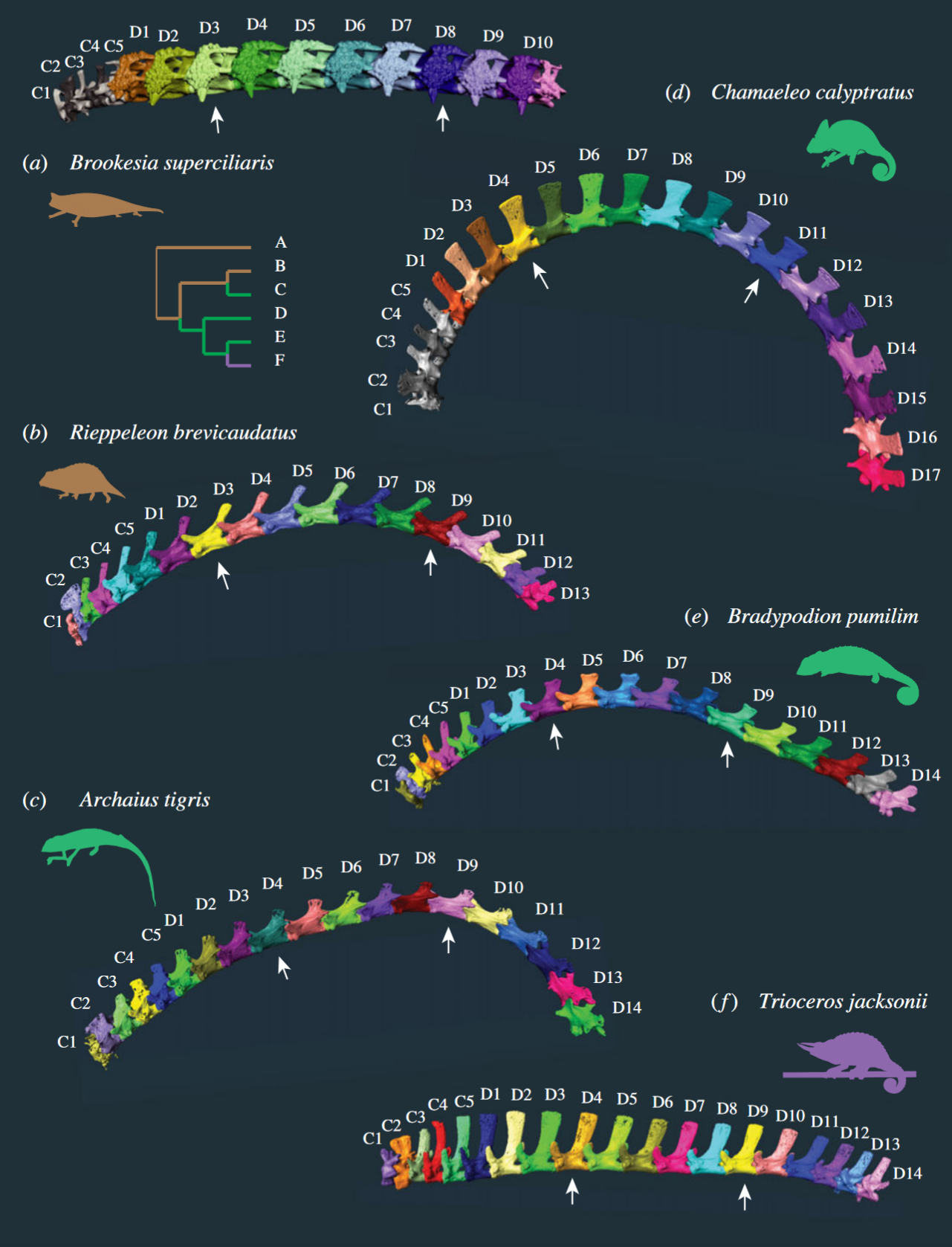
They measured the already existing CT scans on Morphosource.org of a total of 28 chameleons of different species. Brookesia perarmata, Brookesia superciliaris, Brookesia thieli, Palleon nasus, Rhampholeon platyceps, Rhampholeon spectrum, Rieppeleon brevicaudatus and Rieppeleon kerstenii were classified as ground dwellers. Archaius tigris, Bradypodion melanocephalum, Bradypodion pumilum, Bradypodion thamnobates, Calumma amber, Calumma brevicorne, Calumma parsonii, Chamaeleo calyptratus, Chamaeleo gracilis, hamaeleo zeylanicus, Furcifer lateralis, Furcifer pardalis, Furcifer verrucosus, Kinyongia carpenteri, Kinyongia tavetana, Kinyongia xenorhina, Nadzikambia mlanjensis, Trioceros feae, Trioceros jacksonii and Trioceros quadricornis were considered arboreal. The vertebrae were counted and the width of the lamina, length, width, height of the vertebral body, and the height of the spinous process and transverse processes on each vertebra were measured. In addition, the so-called prezygapophysial angle was determined. This is the angle of the intervertebral joint, i.e. the contact surfaces between the individual vertebrae. The measurements of ground and tree dwellers were compared and statistically evaluated. Only the vertebral column of the trunk was considered, the caudal vertebral column was left out.

First of all, the results showed that ground-dwelling chameleons generally have fewer trunk vertebrae (15 to 19) than tree-dwelling chameleons (18 to 23). The trunk spine of almost all species could be divided into the already known three areas: Cervical spine and anterior and posterior dorsal spine. A thoracic and lumbar spine as in mammals is generally not distinguished in chameleons because of the continuous ribs. Five chameleon species had four regions instead of three: they had an anterior and a posterior cervical spine, the anterior one consisting of only two vertebrae with rib processes. Six chameleon species had two additional lumbar vertebrae and one species had three transitional vertebrae in the region between the cervical and dorsal spine. In Kinyongia carpenteri, a total of five regions could be distinguished in the trunk spine: The chameleon had anterior and posterior cervical vertebrae as well as anterior and posterior dorsal vertebrae and two additional lumbar vertebrae. Brookesia perarmata was also a special case: the trunk spine of this chameleon consisted of only two regions and at the same time the smallest number of vertebrae of all species studied.

The greatest differences between ground and tree-dwelling chameleons were found in the prezygapophyseal angle (PZA) and the height of the spinous process. The intervertebral joint surfaces in the anterior dorsal vertebrae of tree-dwelling chameleons were clearly more dorsoventrally oriented and smaller than in ground-dwelling species. Several tree-dwellers showed a PZA of less than 90°. In tree-dwelling chameleons, the largest spinous processes were located at the transition from the cervical to the dorsal spine. Among the ground-dwelling species, the spinous processes were similar only in Palleon nasus. In ground-dwelling chameleons, the appearance of the spinous process varied greatly. Rieppeleon, for example, showed narrow, backward-sloping spinous processes, while the spinous processes in Brookesia were more like a kind of bone bridge than a process. Archaius tigris was an exception: The spinous processes in this chameleon hardly differed along the entire spine.

The authors conclude from the results that the anatomy of the different vertebrae is strongly related to the chameleons’ way of life and different locomotion. The intervertebral joint surfaces in tree-dwelling chameleons are probably important for climbing by supporting the function of the shoulder girdle. Reduced mobility in the mediolateral plane provides greater trunk stiffness, which facilitates climbing in arboreal dwellers. Stiffening of the axial skeleton (skull, trunk spine and thorax) is also known from tree-dwelling mammals. The larger spinous processes in larger chameleons could facilitate shoulder girdle rotation and muscle movement, resulting in increased stride length, better head support, and thus possibly easier feeding.

Morphological and functional regionalization of trunk vertebrae as an adaption for arboreal locomotion in chameleons
Julia Molnar, Akinobu Watanabe
Royal Society Open Science 10, 2023: 221509
DOI: 10.1098/rsos.221509

Illustration: Spines of different chameleon species

Long-term study on sperm collection in chameleons

Long-term study on sperm collection in chameleons

Tiermedizin Science

Assisted reproduction has become increasingly common in the conservation of extremely rare animals such as the Spix’s macaw or northern white rhinoceros in recent years. In reptiles, on the other hand, there have only been a few studies on assisted reproduction, and only a few on chameleons in particular. Scientists from the USA have now conducted a study on male Veiled and Panther Chameleons (Chamaeleo calyptratus and Furcifer pardalis).

At Louisiana State University, 16 males of each species were kept under standardised conditions for over a year. The panther chameleons were purchased from a US breeder, the Yemen chameleons from a dealer who had taken them from the introduced wild chameleon population in Florida. All males were kept individually in ZooMed screen cages, equipped with automatic sprinklers and artificial plants. Temperatures were around 28-29°C during the day with spots to seek higher values. 12 h UV-B irradiation per day was offered. They were fed with crickets and zophobas.

Before the start of the study, all 32 chameleons were clinically examined and parasites were treated. Only after a month of acclimatisation did the actual study begin. During the study year, all chameleons were put under anaesthesia twice a month. Each time, blood was taken from the ventral tail vein or the jugular vein to determine the testosterone concentration. Ultrasound was used to measure the size of the testicles. In addition, each time an attempt was made to obtain sperm by electroejaculation. Electroejaculation involved inserting a small metal probe into the cleaned cloaca. Each chameleon was then treated up to three times in succession with up to 15 electric shocks of 0.1/0.2/0.3 mAs. The semen collection experiments were stopped as soon as the animal ejaculated. The sperm collected was preserved and examined for ejaculate volume, presence of sperm, sperm motility, concentration, and morphology.

The results suggest that Veiled Chameleons follow a so-called prenuptial reproductive strategy under constant husbandry conditions. The testosterone concentration in the blood already increased before the sperm volume of the males had reached its maximum. The months of May, April, and June brought the best sperm volumes, the most sperm was produced by electroejaculations in the third attempt. Testicle sizes also varied throughout the year, with the largest measurements from August to December.

Panther chameleons, on the other hand, seem to follow a postnuptial reproductive strategy. In them, most sperm could only be obtained well after the highest point of testosterone concentration. The electroejaculations worked best in March, April, May and June. Much more often than in Yemen chameleons, electroejaculation in panther chameleons worked already in the first attempt. The size of the testicles also varied throughout the year, but most were largest in the months mentioned above. Together with the factors mentioned above, the volume of ejaculate, sperm concentration, sperm motility and sperm morphology also changed during the year.

The authors recommend that electroejaculation in chameleons should generally only be performed under anaesthesia. The success rate for spermatozoa in the two highest cases was 82 and 88%, which is similar to the success in other reptiles during their reproductive season. The mortality rate among the 32 animals was only 0.12% over the whole year. One panther chameleon died after 10 months during the 20th anaesthesia, after death kidney damage was detected. From the low mortality rate, the authors conclude that electroejaculation rather does not play a role in the development of kidney disease, as was suspected in other studies. However, an examination of the blood for kidney values was not carried out on any of the surviving chameleons after the study. It also remains unclear what role the lack of imitation of rainy and dry seasons during the year plays for both species and their reproductive cycle.

Characterizing the annual reproductive cycles of captive male veiled chameleons (Chamaeleo calyptratus) and panther chameleons (Furcifer pardalis)
Sean M. Perry, Sarah R. Camlic, Ian Konsker, Michael Lierz, Mark A. Mitchell
Journal of Herpetological Medicine and Surgery 33 (1), 2023, pp. 45-60
DOI: 10.5818/JHMS-D-22-00037

Unusual parasite discovered in Furcifer campani

Unusual parasite discovered in Furcifer campani

Tiermedizin Science

Physiologists, microbiologists and veterinarians from the USA recently described an unusual case of a parasite infestation in Furcifer campani. This is probably the new discovery of an as-yet-undescribed chameleon parasite.

In 2021, 11 Furcifer campani had been imported from Madagascar as wild-caught specimens and were kept privately. Two months after importation, unusual behaviour was noticed in one male and one female of the group. The two animals basked in the sun for unusually long periods of time, specifically seeking out temperatures of 29-30°C as well as places with enormously high UV indices compared to the other chameleons. Within the next three months, both Furcifer campani visibly lost weight, although the food supply was increased and a good food intake could be observed. At the same time, a lighter skin colouration was noticed. Faecal examinations by flotation were negative. Finally, both chameleons became lethargic, and closed their eyes during the day. A bloated abdomen and increased watery faeces were observed. Both Furcifer campani died.

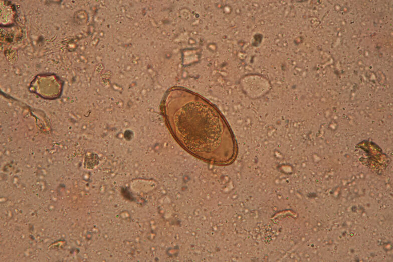
Histological examination confirmed muscle atrophy and cachexia. Massive infiltration of the liver and gastrointestinal tract with large amounts of spores could be detected in both chameleons. The spores proved positive in Grocott’s methenamine-based silver stain and the PAS stain. Morphologically, the spores were classified as Dermocystidium-like. Investigations by PCR revealed a high similarity with Dermocystidium salmonis, but the exact pathogen could not be determined with certainty.

The genus Dermocystidum is a parasitic microorganism that is classified as a protist (it is neither a fungus nor an animal or plant). It is interesting that they have so far been known mainly from fish and amphibians, occasionally also from mammals. So far, not a single case of infestation with Dermocystidium has been described from reptiles. It could therefore be an undescribed, new species that is possibly even chameleon-specific. Effective therapy is not yet known.

A unique disease presentation associated with a mesomycetozoean-like organism in the jeweled chameleon (Furcifer campani)
Michael Nash, Emily A. McDermott, Ashley K. McGrew, Juan Muñoz, Dayna Willems
Journal of Herpetological Medicine and Surgery, February 2023
DOI: 10.5818/JHMS-D-22-00033

Minimally invasive methods for obtaining DNA samples from chameleons

Minimally invasive methods for obtaining DNA samples from chameleons

Tiermedizin Science

To reliably identify or compare chameleon species, genetic samples of the animals concerned are necessary. Traditionally, scientists have used organ or muscle samples from euthanized chameleons in museum collections or – less commonly – cut tail tips or blood samples from living chameleons. Researchers at the American College in Athens, Greece, have studied whether more minimally invasive methods would also be a good alternative.

They sampled 23 Chamaeleo africanus in the area of the lagoon of Pylos (Divari wetland between Gialova and the bay of Voidokilia) in the Peloponnese in Greece using buccal swabs. This involves running a sterile swab on the inside of the cheek through the chameleon’s mouth for six seconds. Blood was taken from the ventral tail vein of eight other Chamaeleo africanus for comparison. Sampling took less than a minute. Afterward, the chameleons were returned to where they were found. The swabs were transported refrigerated in a special buffer solution in Eppendorf cups and then frozen.

In the laboratory, the researchers were able to extract both nuclear and mitochondrial DNA from all the swabs. However, the quantity and quality of the DNA extracted were lower than in the blood samples. For most applications such as PCR amplification and gene sequencing, however, the scientists said the quantity was sufficient. In terms of invasiveness and destructiveness, buccal swabbing is certainly preferable to killing or injuring individual chameleons. Studies on other reptiles suggest that rapid freezing is not mandatory either – in the field, a functioning cool chain could become a problem in many chameleons’ countries of origin. The current study advises against ethanol as a fixing solution; the buffer solutions used lead to better results.

Buccal swabbing appears to be less applicable for cases where additional material for future studies might be preserved, for example when describing new species, or when sequencing the entire genome. However, the method is certainly a good alternative, especially for particularly small chameleon populations where lethal sampling could already significantly limit the breeding pool.

Buccal swabs as an effective alternative to traditional tissue sampling methods for DNA analyses in Chamaeleonidae
Maria Koutsokali, Christina Dianni and Michael Valahas
Wildlife Biology
DOI: 10.1002/wlb3.01052