Histological examinations of organ tissue are part of every pathological examination in veterinary medicine. They are also frequently carried out in reptiles, but there are few studies on the histology of healthy organ tissue. An Arabic publication now deals with histological sections of chameleon livers.
Seven adult Yemen chameleons were captured in Abha City in the Aseer region and then killed with ether inhalation. The livers were placed in formalin and then poured into paraffin to make sections.
Morphologically, the liver was found to be a two-lobed, dark brown organ approximately 3.7 x 2 cm in size, which lies in the coelomic cavity in front of the stomach and surrounds the gall bladder. As in other animals, a capsule of connective tissue surrounds the liver.
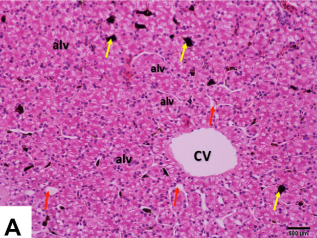
Histologically, the liver of Yemen chameleons resembles that of other vertebrates in many respects. The liver capsule consists of closely spaced collagenous fibres and smooth muscle fibres. Normally, trabecular connective tissue divides the liver itself into many small lobules, but such a structure does not appear to be present in Yemen chameleons. In contrast to mammals, the liver cells (hepatocytes) are not arranged radially around a vein, but rather irregularly in follicles or alveoli. The hepatocytes are surrounded by capillary blood vessels. So-called melanoma macrophages, which are not found in birds and mammals, can be seen in the blood vessels. The hepatocytes in the Yemen chameleon are polyhedral or pyramid-shaped and usually contain several large, round cell nuclei in the periphery. The nuclei contain conspicuously dark nucleoli. Occasionally nuclei are central. Under haematoxylin-eosin (HE) staining, the hepatocytes appear very eosinophilic. In the connective tissue, branches of the portal vein, hepatic artery, small bile ducts and lymphatic vessels could be visualised. Haematopoietic tissue was found in the area directly under the liver capsule.
In addition to the histological examination, several pieces of liver were also examined using transmission electron microscopy. Images of both examination methods can be found in the publication.
Histomorphological, histochemical and ultrastructural studies on the healthy liver of Yemen Veiled Chameleon (Chamaeleo calyptratus) in Southern Saudi Arabia
Amin A. Al-Doaiss, Mohammed A. Alshehri, Ali A. Shati, Mohammad Y. Alfaifi, Mohammed A. Al-Kahtani, Ahmed Ezzat Ahmed, Refaat A. Eid, Laila A. Al-Shuraym, Fahd A. Al-Mekhlafi, Mohammed Al Zahrani, Mohammed Mubarak
International Journal of Morphology 41(5), 2023: pp. 1513-1526.
DOI: none
Image: Histological section of the liver of a Yemen chameleon from the above-mentioned publication


