Scientists discover second ‘super muscle’ in chameleon tongues

Scientists discover second ‘super muscle’ in chameleon tongues

Science

Scientists have been investigating how chameleons’ tongues can shoot out of their mouths to such an extreme length for as long as anyone can remember. US biologists have now been able to solve another piece of the chameleon tongue puzzle.

In the study, experiments were carried out on 15 Veiled chameleons (Chamaeleo calyptratus). All 15 animals were first anaesthetised using overdosed gas anaesthesia and then killed by decapitation. The four tongue muscles were then dissected out and clamped in measuring devices. The muscles were stimulated with an electric current and the passive/active forces and various muscle lengths were measured. A series of calculations were then carried out and muscles were examined histologically. In addition, embryos previously obtained from eggs were analysed immunohistochemically to determine whether the tongue muscles develop from the same or two different muscle systems.

The results of the study are very interesting and focus primarily on the so-called sarcomeres of the muscles. A sarcomere is the smallest contractile unit of the muscle – i.e. the part that is responsible for the expansion and contraction of the muscles. At both ends of each sarcomere are the so-called intermediate discs (German “Zwischenscheibe”), abbreviated as Z-discs. Transverse striated muscles, i.e. the muscles that move the arms, legs and trunk of a vertebrate, can shorten to about half their resting length. In chameleons, however, there is a very special type of muscle, the so-called supercontracting muscle. By definition, this is a striated muscle that is able to shorten to less than half its resting length. The rectractor of the tongue, the hyoglossus muscle, is just such a muscle. In this muscle, perforated Z-discs on the sarcomeres ensure that it can stretch far better than normal striated muscles.

In the present study, it was found that a second supercontracting muscle is involved in the tongue shot: the sternohyoid superficialis muscle. In its counterpart, the sternohyoid profundus muscle, surprisingly, no perforated Z-discs could be detected at the sarcomeres. However, its length-tension ratio corresponded to the two supercontracting tongue muscles. This could be compensated for by the very broad attachment of the muscle to the hyoid bone. During tongue shooting, these areas of the hyoid bone are rapidly rotated, which could mechanically alter the sarcomeres.

Using immunohistochemistry, the biologists were also able to show in chameleon embryos that the two muscles develop from different origins, which is consistent with the different sarcomeres. Both the hyoglossus muscle and the sternohyoid muscles form a muscular unit, with one of the muscles even extending to the sternum. This means that the maximum length of the chameleon tongue when shooting is not only made possible by the special properties of the tongue muscles, but also by the overall length of the muscular unit. No other vertebrate in the world has ever been found to have two supercontracting muscles.

Feats of supercontractile strength: functional convergence of supercontracting muscle properties among hyoid musculature in chameleons
Nikole G. Schneider, Nicholas A. Henchal, Raul E. Diaz Jr., Christopher V. Anderson
Proceedings B of Royal Society Publishing, 2025
DOI: 10.1098/rspb.2025.0078

Figure: Schematic representation of the tongue muscles and hyoid bone in the Veiled chameleon from the aforementioned publication

Veiled chameleon distributed over 12 new counties in Florida (USA)

Veiled chameleon distributed over 12 new counties in Florida (USA)

Verbreitung Science

Introduced Veiled chameleons (Chamaeleo calyptratus) have been living in Florida (USA) since at least 2002. The first wild Veiled chameleons were found in Collier County, two years later animals were observed in Fort Myers in Lee County. This was followed by findings in Hendry, Miamia-Dade, Broward, St. Lucie, Palm Beach, Monroe, Alachua and Hillsborough County. Now an author in the Herpetological Review reports on 12 further populations in Florida: in Brevard, Charlotte, De Soto, Glades, Indian River, Lake, Manatee, Osceola, Pinellas, Polk, Sarasota and Seminole County.

He used data from iNaturalist and EDDMapS. The author suspects that most of the new finds could be due to so-called chameleon ranching. Chameleons are deliberately released into other habitats in order to later collect and sell the resulting juveniles. But even without prior release, collecting (‘harvesting’) animals for sale has become a source of income in Florida, which has also led to the fact that distribution data on new populations is rarely published. It is now a common recreational activity in Florida to search for Veiled chameleons at night. There are even commercial operators offering guided tours.

One problem is increasingly the owners of private property who feel disturbed by ‘chameleon tourism’. The impact on native wildlife in the USA is still unclear. Theoretically, Veiled chameleons could eat smaller mammals or young birds, but there have been no reports of such incidents in Florida to date.

It seems increasingly unlikely that it will be possible to get rid of the introduced Veiled chameleons. A one-year trial in Lake Worth Beach (Palm Beach) resulted in 1043 chameleons being caught during 71 collection campaigns, but not in the elimination of the population living there.

New County Records for the Veiled Chameleon (Chamaeleo calyptratus) in Florida, USA
Kevin M. Enge
Herpetological Review 55 (2), 2025: 223-226.
DOI: not available

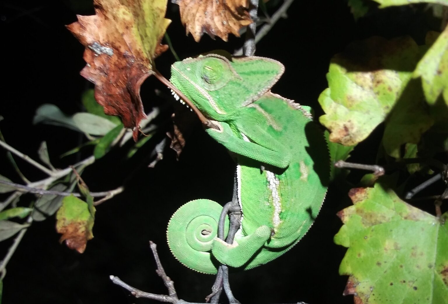
Photo: Chamaeleo calyptratus, found and photographed in Fort Myers (USA) by Andrew Durso, CC-BY

Interested parties wanted for breeding project with Chamaeleo calyptratus calcarifer

Interested parties wanted for breeding project with Chamaeleo calyptratus calcarifer

Abzugeben Projects

The larger subspecies of the Veiled Chameleon, Chamaeleo calyptratus calcarifer, from the Tihama on the Arabian Peninsula has received little attention in the herpetoculture hobby in recent years. It is therefore not surprising that notes on this subspecies are not particularly frequent in the current literature and that there is also a lack of husbandry and breeding reports. Sophie Obermaier, a member of AG Chamäleons from Berlin, would like to change this and breed the species in the long term. She herself has several adults of the species and already has over 50 eggs that are currently being incubated. Anyone who has had experience with the subspecies, would like to participate by keeping a few animals themselves or would like to take on young animals for scientific work is cordially invited to contact Sophie by email at chartar.0702@gmail.com.

(Deutsch) Jemenchamäleons suchen neue Halter

(Deutsch) Jemenchamäleons suchen neue Halter

Abzugeben

Two Veiled Chameleons arrived at the reptile rescue centre in Munich yesterday. Both animals were confiscated and will not be returned to their original owners, so they will probably be released for rehoming shortly. They are two adult females who so far appear to be healthy. The results of the blood check-up and the quarantine at the rescue centre are still pending. However, anyone who is already interested in the two or one of the females is welcome to contact the rescue centre now. A questionnaire for interested parties can be found on their website.

Photo: One of the female Veiled Chameleons, photographed at the reptile rescue centre, Munich

Sex chromosomes in chameleons

Sex chromosomes in chameleons

Science

Which sex chromosomes are present in chameleons has so far been studied rather sparsely. The Madagascan chameleon genus Furcifer is known to have Z and W chromosomes, although sometimes several Z chromosomes occur, so-called neo-sex chromosomes. Recently in the Czech Republic, scientists examined this deeper.

Blood and tissue samples were taken from 13 chameleons to isolate DNA. The animals sampled included one male and one female each of the species Brookesia therezieni, Calumma glawi, Calumma parsonii, Chamaeleo calyptratus, Furcifer campani, Furcifer labordi, Furcifer lateralis, Furcifer oustaleti, Furcifer pardalis, Furcifer rhinoceratus, Furcifer viridis, Kinyongia boehmei and Trioceros johnstoni. Only in Furcifer oustaleti were two females sampled. Subsequently, the Z1 chromosomes of the panther chameleons and the Z and W chromosomes were analysed by microdissection. Gene coverage analyses were performed for carpet and panther chameleons. In addition, qPCRs were performed to compare the homology of the Z chromosomes.

The results show that the morphology of the Z1 chromosomes of panther chameleons corresponds to the Z chromosome of the entire genus Furcifer. The Z1 chromosome of panther chameleons thus corresponds to the Z chromosome of Furcifer oustaleti. The Z2 chromosome of panther chameleons, on the other hand, is a neo-sex chromosome. Both the Z and W chromosomes in Furcifer oustaleti are probably pseudautosomal. 42 genes have been described as specific for the W chromosome.

A total of 16,947 genes were identified in Furcifer lateralis and 16,909 genes in Furcifer pardalis. The ratio of the number of genes between females and males is 0.35 and 0.65 for the two species. In panther and carpet chameleons, most of the genes on the W and Z chromosomes were found to be the same, with relatively few genes found only on the W chromosome. This finding is surprising, as the researchers had actually expected that the heterochromatic W in Furcifer species would have lost most of its genes compared to the Z chromosome.

The sex chromosomes of the genus Furcifer probably evolved at least 20 million years ago, which roughly corresponds to the time when the species Furcifer campani split off from the other Furcifer species.

Heteromorphic ZZ/ZW sex chromosomes sharing gene content with mammalian XX/XY are conserved in Madagascan chameleons of the genus Furcifer
Michail Rovatsos, Sofia Mazzoleni, Barbora Augstenová, Marie Altmanová, Petr Velenský, Frank Glaw, Antonio Sanchez, Lukáš Kratochvíl
Scientific Reports 14, 2024: 4898.
DOI: 10.1038/s41598-024-55431-9

New case reports on hemipenes amputation

New case reports on hemipenes amputation

Tiermedizin

The University of Sofia (Bulgaria) has published a new paper with several case reports involving chameleons. The authors describe 16 cases of different lizards that suffered a hemipenis prolapse and their treatment.

The lizards included a panther chameleon (Furcifer pardalis) and two Veiled Chameleons (Chamaeleo calyptratus). All three patients were presented to the veterinarians with bilateral hemipenes prolapse. Initially, the prolapses were bathed in 20% dextrose solution, after which the hemipenes were manually repositioned. However, the prolapses then recurred, so surgery was the final solution. Under general and local anaesthesia administered intramuscularly, the hemipenes were removed, the wound sutured and the remaining small stump repositioned in the respective hemipenes pocket. Meloxicam was administered as an analgesic once a day for 5 days after the operation. Only lizards in which the surgical field appeared to be dying off during the follow-up examinations were given antibiotics for 10 days.

Hemipenectomy in leopard geckos, chameleons and bearded dragons
Seven Mustafa & Iliana Ruzhanova-Gospodinova
Tradition and Modernity in Veterinary Medicine, 2024
DOI: nicht vorhanden

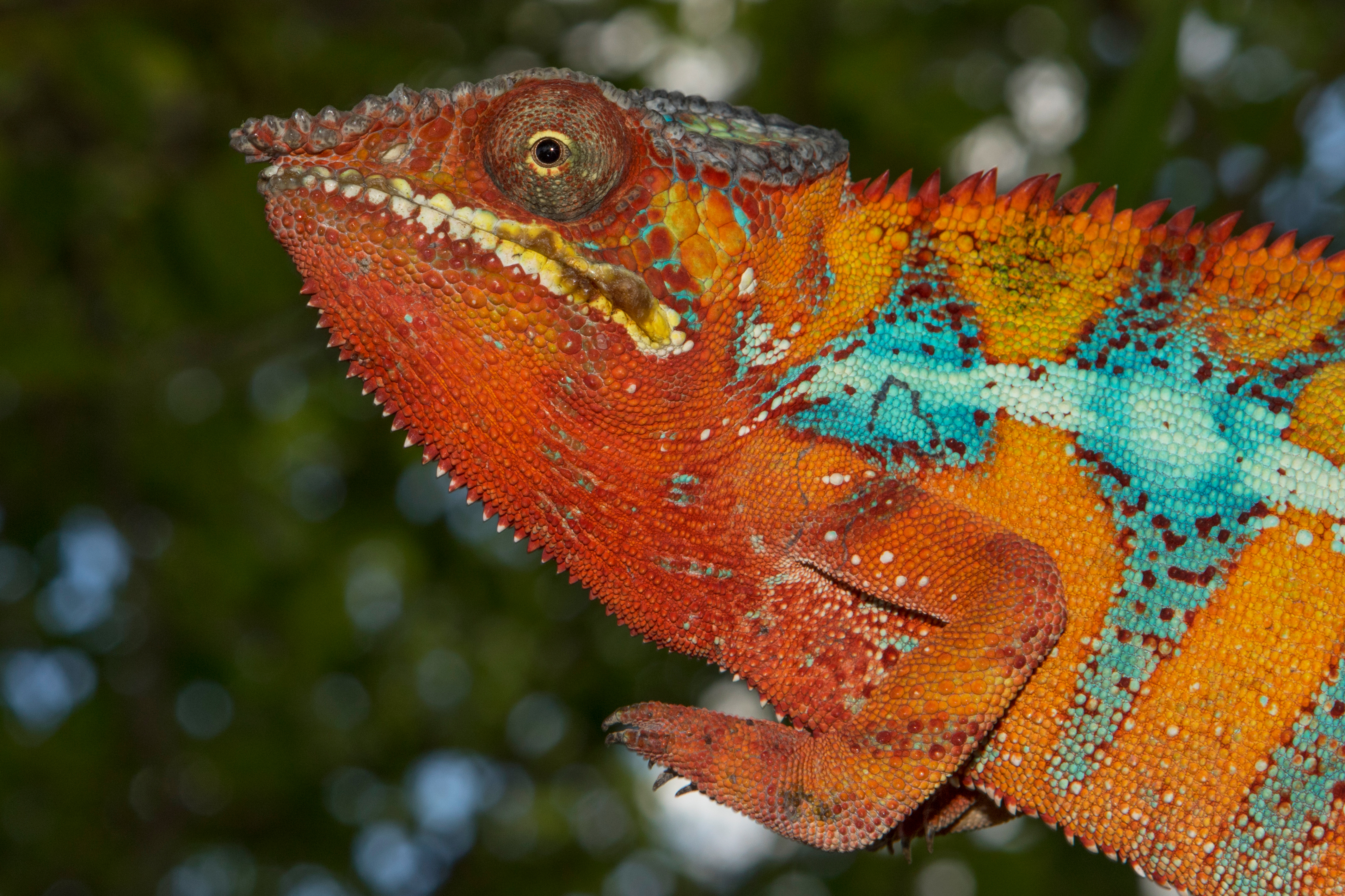
Photo: Panther chameleon, photographed by Alex Laube in Madagascar

Histology of the chameleon liver

Histology of the chameleon liver

Tiermedizin Science

Histological examinations of organ tissue are part of every pathological examination in veterinary medicine. They are also frequently carried out in reptiles, but there are few studies on the histology of healthy organ tissue. An Arabic publication now deals with histological sections of chameleon livers.

Seven adult Yemen chameleons were captured in Abha City in the Aseer region and then killed with ether inhalation. The livers were placed in formalin and then poured into paraffin to make sections.

Morphologically, the liver was found to be a two-lobed, dark brown organ approximately 3.7 x 2 cm in size, which lies in the coelomic cavity in front of the stomach and surrounds the gall bladder. As in other animals, a capsule of connective tissue surrounds the liver.

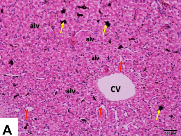
Histologically, the liver of Yemen chameleons resembles that of other vertebrates in many respects. The liver capsule consists of closely spaced collagenous fibres and smooth muscle fibres. Normally, trabecular connective tissue divides the liver itself into many small lobules, but such a structure does not appear to be present in Yemen chameleons. In contrast to mammals, the liver cells (hepatocytes) are not arranged radially around a vein, but rather irregularly in follicles or alveoli. The hepatocytes are surrounded by capillary blood vessels. So-called melanoma macrophages, which are not found in birds and mammals, can be seen in the blood vessels. The hepatocytes in the Yemen chameleon are polyhedral or pyramid-shaped and usually contain several large, round cell nuclei in the periphery. The nuclei contain conspicuously dark nucleoli. Occasionally nuclei are central. Under haematoxylin-eosin (HE) staining, the hepatocytes appear very eosinophilic. In the connective tissue, branches of the portal vein, hepatic artery, small bile ducts and lymphatic vessels could be visualised. Haematopoietic tissue was found in the area directly under the liver capsule.

In addition to the histological examination, several pieces of liver were also examined using transmission electron microscopy. Images of both examination methods can be found in the publication.

Histomorphological, histochemical and ultrastructural studies on the healthy liver of Yemen Veiled Chameleon (Chamaeleo calyptratus) in Southern Saudi Arabia
Amin A. Al-Doaiss, Mohammed A. Alshehri, Ali A. Shati, Mohammad Y. Alfaifi, Mohammed A. Al-Kahtani, Ahmed Ezzat Ahmed, Refaat A. Eid, Laila A. Al-Shuraym, Fahd A. Al-Mekhlafi, Mohammed Al Zahrani, Mohammed Mubarak
International Journal of Morphology 41(5), 2023: pp. 1513-1526.
DOI: none

Image: Histological section of the liver of a Yemen chameleon from the above-mentioned publication

Zoonotic potential of Yemen chameleons in Gran Canaria (Spain)

Zoonotic potential of Yemen chameleons in Gran Canaria (Spain)

Science

The Canary Islands are located northwest of Africa near the coast of Morocco. On Gran Canaria, the second largest island, around 290 of over 1000 plant and animal species have been introduced, i.e. species that do not originally occur there. Since at least 2017, there have been free-living Veiled chameleons (Chamaeleo calyptratus) on Gran Canaria. Spanish scientists have now investigated whether this introduced chameleon population could have zoonotic potential.

They examined 40 Veiled chameleons that had previously been caught and killed by Red de Alerta Temprana de Canarias para la Detección e Intervención de Especies Exóticas Invasoras in Arucas. 36 of the chameleons were adults, four were juveniles. Intestinal contents were taken from each chameleon and analysed for the presence of different bacteria using various methods.

At least one of the bacteria sought was found in 28 of the Veiled chameleons. About half of the chameleons had Yersinia enterocolitica, which is the highest prevalence ever recorded for this bacterium in reptiles. The bacterium can cause diarrhoea in humans, among other things. It is unclear how the Veiled chameleons became infected with it – possibly via insects. 16 of the Veiled chameleons had salmonella in their intestines. Salmonella is very common in reptiles and has even been found in endemic species on Gran Canaria. Pseudomonas is also frequently found in reptiles and was detected in the intestines of 13 animals. Two Veiled chameleons were infected with Campylobacter, in one of them Campylobacter lari could be identified. This bacterium can rarely cause illness in humans, but no pathogenic potential for humans is known for the species otherwise common in reptiles. Campylobacter lari has so far been detected mainly in seafood and birds – it is possible that the Veiled Chameleons picked up the bacterium on the coast and did not bring it with them. Three Veiled chameleons had Escherichia coli, which in rare cases can lead to haemolytic uraemic syndrome (HUS) in humans. Another two chameleons had Listeria monocytogenes in their intestines, which can be dangerous for pregnant women if ingested with food. Five Veiled Chameleons had mycobacteria, several of which were found to be non-tuberculous infections. Staphylococci were detected in seven chameleons, but they are part of the normal skin flora. However, five isolates were positive for resistance to certain antibiotics, which is becoming an increasing problem with Staphylococcus aureus in humans. Most recently, Vibrio was detected in a single Yemen chameleon, some species of which can cause diarrhoea in humans. The bacterium has previously been detected in introduced anoles on Tenerife.

The authors state that there is a zoonotic potential for humans due to the handling of introduced Veiled Chameleons on Gran Canaria. However, the extent to which there is a real risk for humans as well as endemic species needs to be further investigated.

Study of zoonotic pathogens in alien population of Veiled Chameleons (Chamaeleo calyptratus) in the Canary Islands (Spain)
Román Pino-Vera, Néstor Abreu-Acosta, Pilar Foronda
Animals 13 (14), 2023
DOI:  10.3390/ani13142288

Twins in Veiled Chameleons in Latvia

Twins in Veiled Chameleons in Latvia

Short messages Nachzuchten

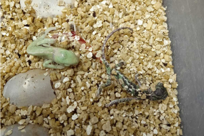
The hatching of two pairs of twins of Veiled Chameleons (Chamaeleo calyptratus) in Latvia has been reported from Riga. The parents live at Riga Zoo and the young hatched in March 2022. The twins were two male and two female Veiled Chameleons, each pair in one egg. They came from a clutch of 85 eggs, of which 48 eventually hatched. All four young animals were active at first and accepted food. At the age of two months, one of the juveniles died, the remaining three were still alive in February 2023.

The article also gives a brief overview of cases of twins in reptiles from the existing literature.

A review of twinning in lizards and a report of Veiled Chameleon (Chamaeleo calyptratus) twin births
Alessandro di Marzio, Elza Birbele, Lucia Puchades, Andris Lazdiņš
Herpetology Notes 16: 471-476, 2023
DOI:

Photo: One of the twin pairs at hatching